Uterine fibroids: what are they, types, diagnosis and treatment

- What are uterine fibroids?
- Location of uterine fibroids
- What types of uterine fibroid can we find?
- What is the origin of fibroids?
- What symptoms do women with uterine fibroids suffer from?
- How are uterine fibroids diagnosed?
- Treatment of uterine fibroids
What are uterine fibroids?
- Uterine fibroids are abnormal growths of tissue in the muscular wall of the uterus forming solid masses of different sizes.
- There are several factors thought to be related to the development of uterine fibroids.
- The definitive treatment for uterine fibroids is hysterectomy, although there are other types of minor surgery.
- Uterine fibroids are proliferations of cells of the muscular wall of the uterus forming solid masses that can vary in size and position inside the uterus. They are the most common tumours of the pelvic cavity in women.
Uterine fibroids are proliferations of cells in the muscular wall of the uterus forming solid masses that can vary in size and position inside the uterus. They are the most common tumours of the pelvic cavity in women.

Do you need myomectomy surgery?
Request a free and immediate appointment with our specialists
It is estimated that they are present, although without symptoms, in about 20-40% of women of reproductive age, increasing to 70% in women over 70 years of age.
Of all women with uterine fibroids, only 20-25% have accompanying symptoms. They are also known as uterine leiomyomatosis, fibromyomas, leiofibromyomas or fibroleiomyomas.
Location of uterine fibroids
Uterine fibroids can occur in almost any location in the uterus. They are usually confined to one of the 3 layers of tissue that make up the uterus called the myometrium.

The uterus is the largest organ of the female reproductive organs, located inside the pelvic cavity between the bladder and the rectum. It is made up of 3 layers, if we look at them from the outside in:
- The first layer is the peritoneum (layer of tissue that surrounds the abdominal cavity and surrounds the uterus laterally and posteriorly),
- The second is the myometrium (made up of muscle cells and collagen).
- The last is the endometrium or mucosa (a serous and glandular layer that is responsible for proliferating under the influence of hormones during the various phases of a woman's ovarian cycle and where the foetus will implant during pregnancy).
What types of uterine fibroid can we find?
There is a great variety of possibilities to typify the different myomas due to the multitude of possibilities and classifications according to their characteristics. If we base our classification on their location we can find:
- Intramural uterine fibroids, which are fibroids that develop in the muscular wall of the uterus.
- Submucosal fibroids are those that are under the mucosa, the inner surface of the uterus.
- Subserosal myomas are those found under the serosal layer, which is the superficial layer of the uterus.
Both submucosal and subserosal fibroids can be pedunculated, meaning that they detach from the uterus and are attached to it only by a small stalk.

Do you need myomectomy surgery?
Request a free and immediate appointment with our specialists
If we take into account their microscopic characteristics of cell composition and other different alterations, we can classify myomas into: cellular uterine myoma, epitheloid uterine myoma, myxoid uterine myoma, symplastic uterine myoma, etc.
What is the origin of fibroids?
Uterine fibroids do not have a specific origin or alteration from which we can ensure the presence and proliferation of uterine fibroids in women. It is considered to be a combination of different risk factors that cause their formation.
There is an evident genetic component after studying mothers and daughters and verifying the increased frequency of both with respect to those women without a family history of uterine fibroids, it seems clear that there is an alteration in one of the genes involved in the proliferation of the muscle cells of the uterus that form one of its layers called the myometrium.
This altered gene is called HMA2. Recently, another altered gene on chromosome 6 has been found to be associated with an increased likelihood of uterine fibroids, the HMGA1 gene.

The hormonal factor is a basic component in the proliferation and formation of uterine fibroids. The endometrial tissue that forms the innermost layer of the uterus has a large number of oestrogen and hormone receptors. This explains why the presence of uterine fibroids before puberty is very low, proliferating their formation and growth at fertile ages and promoting a decrease or atrophy of uterine fibroids after menopause.
Therefore, the hormonal relationship with the formation and increase of uterine fibroids can be classified as linear and direct, being a possible therapeutic target, as we will see below.
According to studies in recent years, it seems clear that the formation of these solid tumours of the uterus, called uterine fibroids, is due to the proliferation of a single myometrial cell that increases in size and divides exponentially with respect to the others.
The main risk factors predisposing women to uterine myomatosis are:
- Age and number of pregnancies (it has been shown that the younger the age at menarche and the fewer the number of pregnancies, the higher the risk of uterine fibroids).
- Ethnicity (more common in blacks than in other races).
- Use of hormonal contraception (indirect relationship in which the more years of contraceptive use, the lower the risk of uterine myoma).
- Weight, diet and exercise are also risk factors for the presence of uterine fibroids.
- Body Mass Index >30 (obesity) has a 1.5 times higher risk of uterine fibroids.
- Women who do moderate exercise have a lower rate of fibroids than sedentary women.
- Pregnancy seems to be a condition that predisposes women to the formation of new uterine fibroids and the proliferation of existing ones.
- The relationship of lesions in the uterus due to previous interventions or previous births seems to have a direct relationship with the appearance of uterine fibroids at the site of uterine lesions.
- Tobacco decreases the presence of uterine fibroids by decreasing oestrogen formation and hindering the division of myometrial cells. Nevertheless, tobacco is absolutely harmful.
What symptoms do women with uterine fibroids suffer from?
Only 20-25% of women with uterine fibroids ever experience any symptoms that may result from the presence of these uterine alterations. The main symptom, as well as the most frequent, is abnormal uterine bleeding. The frequency and quantity of bleeding is proportional to the number of fibroids, their size, their location and the possible degeneration that may appear in each one of them.
Generally, as we have explained above, uterine fibroids have a clear hormonal relationship and it is common that with the onset of the menopause the size and activity of the fibroids decreases and with it the uterine bleeding they cause. Of all the types of uterine fibroids, the ones most associated with abnormal bleeding are intramural fibroids.

Do you need myomectomy surgery?
Request a free and immediate appointment with our specialists
The next most common symptom associated with the presence of uterine fibroids is a feeling of heaviness or compression at the pelvic level. Patients with uterine fibroids often report a feeling of fullness in the lower abdomen with symptoms of difficulty urinating or increased urinary tract infections.
Generally, this situation is due to an overgrowth of the fibroid that can compromise or compress the bladder and urethra, causing this sensation of occupation and generating urinary problems derived from the obstruction of the urinary tract.
It is possible, although in a smaller number of cases, for some people to suffer abdominal pain due to the presence of fibroids. This situation, if it occurs, is due to necrosis or ischaemia of the fibroid or torsion of the pedicle in the pedunculated fibroids mentioned above.

The link between infertility and uterine fibroids is frequent and is found in a moderate percentage of patients suffering from uterine fibroids. A frequency of 5 to 10% of cases has been established. This is due to several causes, the main ones being:
- Difficulty in implantation of the oocyte in the endometrium of the uterus,
- Difficult access of the sperm to the fallopian tubes for conception with the egg.
- Uterine bleeding caused by myoma that hinders the development of the oocyte even if it has been able to implant in the endometrium of the uterus.
However, the presence of uterine fibroids does not exclude the possibility of pregnancy. It is estimated that 8-10% of pregnancies with moderate or large fibroids in the uterus of pregnant women achieve a normal pregnancy and birth.
How are uterine fibroids diagnosed?
Uterine fibroids are usually suspected when the gynaecologist palpates an enlarged uterus, with irregular contours in the woman, abnormal uterine bleeding or fertility problems.
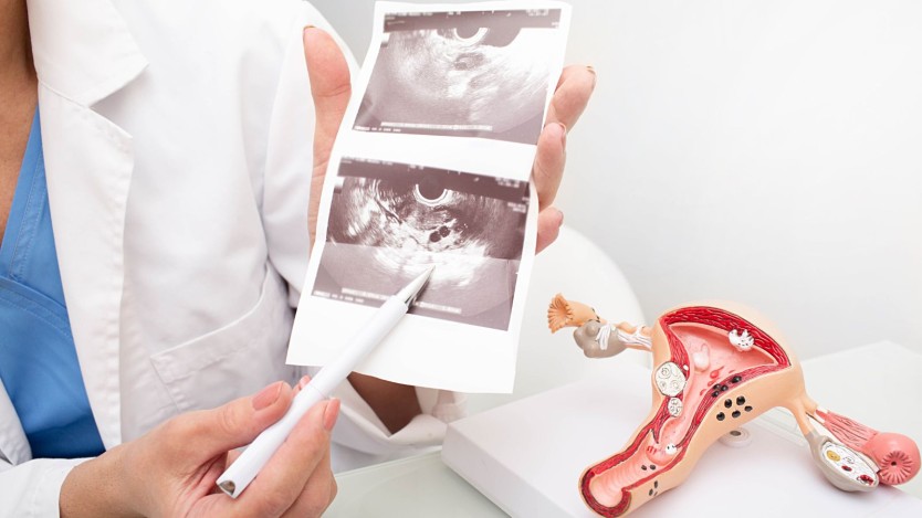
Once the specialist has performed a suspected examination, it is usual to continue the study by performing a vaginal ultrasound, a test that is 95-99% sensitive in expert hands and which usually provides precise information on the size and location of the uterus.

To complete the study it is possible to carry out a hysterosalpingography or a Nuclear Magnetic Resonance Imaging to confirm and detail the anatomical characteristics.
Treatment of uterine fibroids
Definitive treatment of uterine fibroids usually requires surgical removal of the fibroids, especially in women who have not fulfilled their reproductive desire (who have not had children and want to have them).
In patients who are menopausal or who do not wish to become pregnant later, it is possible that certain therapeutic options may be able to partially resolve the symptoms of uterine fibroids without requiring surgery.
Pharmacological treatment of uterine fibroids usually involves the use of hormonal drugs, generally in combination with oestrogens and progestogens or with oestrogen hormone inhibitors.
Generally, the first line of medical action is the administration of progesterone in certain parts of a woman's ovarian cycle. This usually reduces abnormal uterine bleeding, although it has not been shown to reduce the size or number of uterine fibroids.

Another type of drugs with a hormonal component most commonly used are GnRH analogues (gonadotrophin-releasing hormone analogues), this type of mediation seeks to reduce oestrogen production, thereby decreasing the growth and proliferation signals received by the myoma.
Another specific therapeutic group are hormone receptor modulators, substances that compete with oestrogens, progestogens and steroids in the uterus and hinder their function, reducing the proliferation of uterine fibroids.
Finally, the implantation of intrauterine devices with hormonal effect (IUDs) can be associated with a decrease in uterine bleeding and a slight reduction in the size of fibroids.
Surgical intervention is necessary for the definitive resolution of uterine fibroids.
This usually eliminates the fibroid completely and the rate of recurrence of the fibroid.
Of all the surgical techniques, the most effective and the most widely used in women who do not wish to have children is a hysterectomy (surgical technique that consists of the complete removal of the body of the uterus).
In women who wish to have children later or who do not want a complete hysterectomy, myomectomy or removal of uterine fibroids is possible using 3 specific techniques: abdominal myomectomy, laparoscopic myomectomy and myomectomy by hysteroscopy:
- Abdominal myomectomy is the removal of fibroids from inside the uterus by accessing the pelvic cavity through an incision in the abdomen.
- Laparoscopic myomectomy is the removal of uterine fibroids by accessing the pelvic cavity through 2-3 incisions in the abdomen smaller than 4 cm through which surgical instruments in the form of articulated arms smaller than 5 cm can be introduced to remove the fibroids with the least possible abdominal invasion. This technique generally achieves the same results as the abdominal approach with a lower rate of hospitalisation and postoperative complications. This type of intervention is usually performed on large intramural fibroids or fibroids that compress structures adjacent to the uterus such as the rectum or vagina.
- Myomectomy by hysteroscopy consists of the removal of uterine fibroids by accessing the interior of the uterus using a hysteroscope, a surgical device equipped with an optical lens that allows visualisation of the interior of the uterus and the possibility of introducing surgical instruments necessary for the elimination of the fibroids. This type of intervention is usually indicated for submucosal or subserosal fibroids due to their proximity to the endometrium and their limited proliferation towards the outermost tissues of the uterus.

Do you need myomectomy surgery?
Request a free and immediate appointment with our specialists
Medical disclaimer: All the published content in Operarme is intended to disseminate reliable medical information to the general public, and is reviewed by healthcare professionals. In any case should this information be used to perform a diagnosis, indicate a treatment, or replace the medical assessment of a professional in a face to face consultation. Find more information in the links below:
