Intramural uterine fibroids
- What is an intramural uterine fibroid?
- What is the classification of uterine fibroids?
- Symptoms of intramural uterine fibroids
- How is an intramural uterine fibroid diagnosed?
- Surgical treatment for intramural uterine fibroids
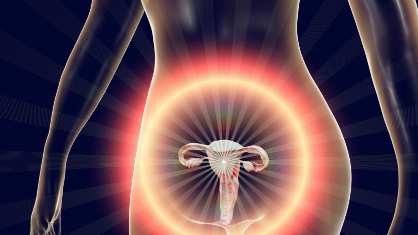
- Intramural uterine fibroids are fibroids that form in the muscular layer of the uterus called the myometrium.
- Intramural uterine fibroids are the most common type of uterine fibroid that can form.
- Treatment of symptomatic intramural uterine fibroids is recommended to be surgical.
What is an intramural uterine fibroid?
An intramural uterine fibroid is a pelvic tumour located within the muscular layers of the uterus. The vast majority of intramural uterine fibroids are benign and form from a single clonal cell (in at least 60% of cases).
The activation of this cell from which the others are generated has a multifactorial and diverse origin that may be due to different causes.
Of all these possible origins in the formation of intramural uterine fibroids, the role of sex hormones such as oestrogens and progestogens seems to be crucial for the formation and growth of this type of fibroids. As a consequence of this situation, one of the possible treatments for intramural uterine fibroids is the use of drugs that modify the concentrations of these sex hormones in order to reduce the size and even achieve the disappearance of small uterine fibroids.

Do you need myomectomy surgery?
Request a free and immediate appointment with our specialists in Gynaecology
What is the classification of uterine fibroids?
Intramural uterine fibroids form part of one of the variants that make up the general entity known as uterine fibroids (also called leiomyomas, fibromyomas or fibroleiomyomas).
From a macroscopic point of view, we can classify four types of uterine fibroids:
- Intramural uterine fibroids: These are uterine fibroids or tumours that are confined within the cells of the muscular wall or myometrium of the uterus. They may be single or multiple and in the latter case may cause dilatation or deformation of the normal architecture of the uterus.
- Subserosal uterine fibroids: These are uterine fibroids or tumours that grow in areas close to the serous layer of the uterus (generally located in the outer third of the uterine wall), generally protruding towards the outside of the uterine cavity in relation to the peritoneum and giving the uterus a bulging appearance.
- Submucosal uterine fibroids: In this case, the fibroids grow close to the endometrial mucosa and protrude into the uterus. These fibroids cause atrophy or destructuring of the uterine endometrial mucosa, causing erosion of the mucosa and moderate or severe bleeding, in addition to the obvious possibility of causing problems in conceiving.
- Pedunculated uterine fibroids: Pedunculated uterine fibroids mainly arise from a submucosal fibroid or a subserosal uterine fibroid. If they form from a submucosal fibroid, they will appear as protrusions towards the interior of the uterine cavity due to uterine contractions, remaining anchored to the uterine wall through a pedicle or isthmus. Generally, this type of fibroid can protrude through the cervix into the vagina and can be seen on the outside in the most severe cases. On the other hand, pedunculated uterine fibroids formed from subserosal fibroids form on the outside of the uterus, remaining suspended in the pelvic cavity.

Symptoms of intramural uterine fibroids
Intramural uterine fibroids, which grow between the muscle fibres of the myometrium of the uterus, are generally the most common cause of infertility problems. This is because they are the ones that reach the largest size and disrupt the usual architecture of the uterine cavity to a greater extent.
The prevalence of infertility has been estimated at around 5-10% of cases of uterine fibroids.

Do you need myomectomy surgery?
Request a free and immediate appointment with our specialists in Gynaecology
In most cases it is caused by alterations in the mucosal contours of the endometrium of the uterus, due to enlargement of the uterine walls that hinder the implantation of the oocyte, altered uterine contractility, making it difficult for the sperm to reach the egg and blocking the entrance to the fallopian tubes, making it difficult for the egg to descend and for the sperm to ascend, thus complicating the union between the two and the formation of the gamete or oocyte.
You can read more information in: Fibroids and pregnancy
However, as with other uterine fibroids, it is possible for intramural uterine fibroids to present any of the symptoms common to all types of uterine fibroids.
The main ones include: abnormal uterine bleeding in the form of menorrhagia or hypermenorrhoea outside the usual menstrual bleeding, pelvic pain and pelvic tumour symptoms due to compression of adjacent structures, mainly urinary symptoms in the form of frequency of urination, acute urinary retention and hydronephrosis or renal involvement in advanced cases.
Compressive symptoms can also affect and provoke intestinal alterations, mainly causing constipation.
How is an intramural uterine fibroid diagnosed?
Intramural uterine fibroids, like other fibroids, are usually diagnosed on the basis of their symptoms, which lead to a clinical suspicion of the presence of uterine fibroids in the woman. Following clinical suspicion, a physical examination is usually performed.
After the physical examination, the next diagnostic test to determine the existence of intramural uterine fibroids is usually transvaginal ultrasound. This type of examination has a sensitivity and specificity of 95% and 99% respectively, which is why it is currently considered the fundamental test for detecting the presence of this type of intramural uterine fibroids.
The rest of the exploratory tests are used to improve the study of the presence of intramural uterine fibroids with a view to a suspected malignancy or to study the disposition of the uterine cavity in greater detail with a view to possible surgical intervention.
Do you need myomectomy surgery?
Request a free and immediate appointment with our specialists in Gynaecology
Within this type of test, hysterosalpingography (injection of contrast into the uterine cavity to visualise the internal architecture of the uterus) gives an idea of the integrity of the fallopian tubes that reach the ovaries and the internal structure of the uterus.
Magnetic resonance imaging or MRI provides information on the possible origin of the myoma and its anatomical relationship with organic structures close to the intramural uterine myoma.

Hysteroscopy (a technique in which an optic is introduced through the vagina into the uterus using a hysteroscope or surgical instrument in the form of a metal rod connected to a video screen) allows direct visualisation of the size, shape and appearance of the myoma and the possibility of taking biopsies to study the anatomopathological composition of the intramural uterine myoma in cases in which it is necessary to resolve doubts about its benignity or malignancy.
Surgical treatment for intramural uterine fibroids
It is normal for a patient suffering from intramural uterine fibroids to also have other types of fibroids, such as subserosal or submucosal fibroids. Depending on the types of uterine fibroids the patient has, their size and location, the surgeon will choose one surgical technique or another to carry out the fibroid removal procedure.
The most commonly used are:
- Hysterectomy: Hysterectomy is the complete removal of the uterus. This option is usually chosen by patients who have already fulfilled their desire to have children or who are very clear that they do not want to have them. In this case, the myomatous uterus is removed, which can be done through an abdominal approach, a laparoscopic approach or through the vagina.
- Hysteroscopy: Hysteroscopy allows the surgeon to remove uterine fibroids without the need for open surgery. Its use is usually reserved for the removal of smaller fibroids. In addition to surgical hysteroscopy, there are diagnostic hysteroscopies that are used to explore the inside of the uterus and determine which type of technique is appropriate. In the removal of uterine fibroids by hysteroscopy, the entire uterus is preserved.
- Myomectomy: The myomectomy operation to remove uterine fibroids allows the symptoms to be resolved without the need to remove the uterus completely. This technique can be performed through an abdominal incision or laparoscopically. The use of one or the other depends on the surgeon's opinion and the severity and size of the fibroids.
Do you need myomectomy surgery?
Request a free and immediate appointment with our specialists in Gynaecology
Medical disclaimer: All the published content in Operarme is intended to disseminate reliable medical information to the general public, and is reviewed by healthcare professionals. In any case should this information be used to perform a diagnosis, indicate a treatment, or replace the medical assessment of a professional in a face to face consultation. Find more information in the links below: